
When talking about Fetal Spina Bifida, a birth defect where the backbone and spinal canal fail to close completely in the womb. Also known as myelomeningocele, it can affect movement, bladder control, and brain development. Understanding this condition early on makes a big difference for families and medical teams.
Fetal spina bifida is a subset of Neural Tube Defects, congenital malformations that arise when the neural tube doesn’t seal properly during the first month of pregnancy. The open tube lets fluid leak, which can damage the spinal cord. Key risk factors include low maternal folate levels, certain genetic variations, and exposure to diabetes‑related sugars. Because the defect forms so early, prevention and detection hinge on nutrition and early prenatal care.
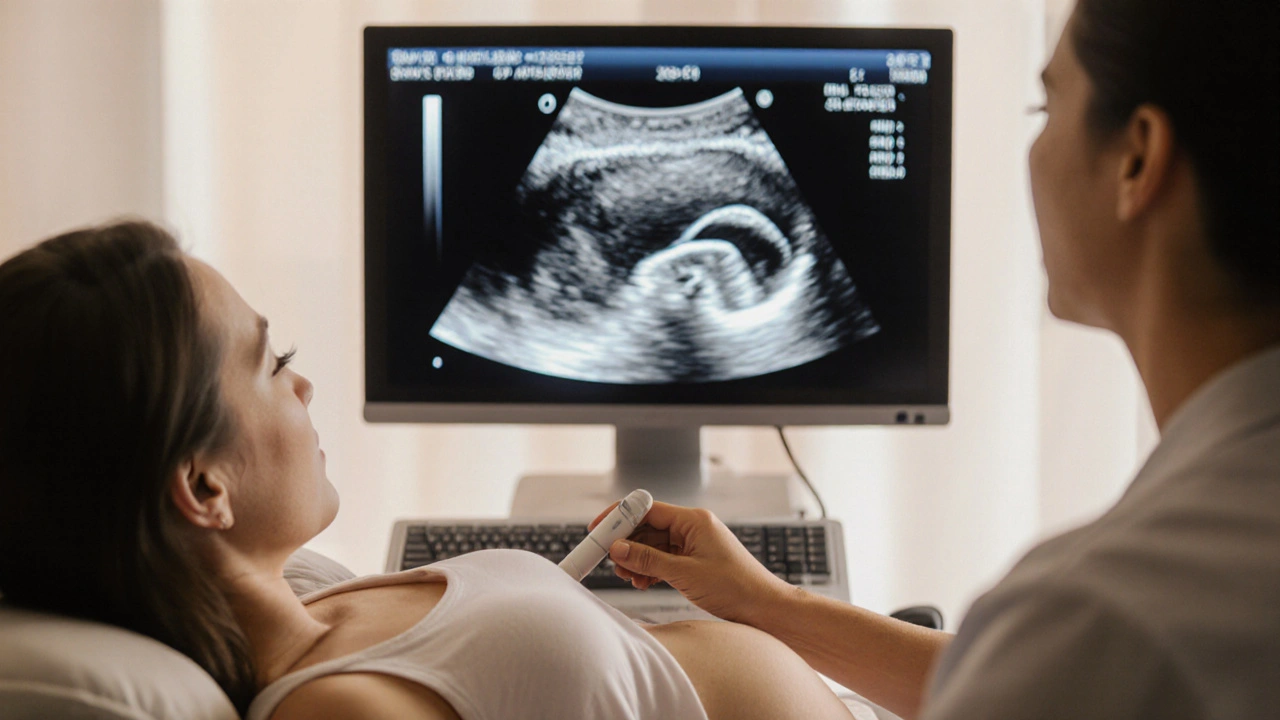
Detecting the defect before birth relies heavily on Prenatal Screening, a set of tests like ultrasound, maternal serum alpha‑fetoprotein (AFP) measurement, and fetal MRI that spot abnormalities in the developing spine. When an elevated AFP result or a suspicious ultrasound appears, doctors usually follow up with a detailed scan around 18‑20 weeks. Early spotting lets families plan for possible fetal surgery or prepare for specialized care after delivery.
Once the condition is confirmed, treatment options split into two main paths: in‑utero surgery and post‑natal repair. Fetal surgery, typically done between 19 and 26 weeks, aims to close the spinal opening before birth, reducing the risk of brain damage caused by fluid leakage. Not every case qualifies—doctors weigh the mother’s health, the baby’s position, and the severity of the defect. After birth, most babies undergo a second operation to reinforce the closure and address any related issues like hydrocephalus.
Outcomes improve dramatically when the defect is fixed early. Babies who receive fetal surgery often show better motor function, fewer shunt placements for hydrocephalus, and higher rates of walking independently. However, surgery carries risks such as preterm labor, so a multidisciplinary team—including obstetricians, neurosurgeons, and neonatologists—must coordinate care before, during, and after the procedure.
Prevention remains the most powerful tool. Health authorities recommend that every woman of child‑bearing age take at least 400 µg of folic acid daily, beginning before conception and continuing through the first trimester. Studies show that this simple supplement can cut the incidence of neural tube defects by up to 70 %. Foods rich in folate—leafy greens, legumes, and fortified cereals—complement the pill and help maintain adequate levels.
Beyond medical steps, families benefit from counseling, support groups, and educational resources. Connecting with other parents who have faced fetal spina bifida can ease anxiety, provide practical tips on managing catheters or mobility aids, and highlight new research breakthroughs. Many hospitals also offer dedicated social workers who can guide insurance navigation and coordinate early intervention services.
Understanding fetal spina bifida gives you a clear view of why early detection, informed prevention, and coordinated treatment matter. Below, you’ll find a curated selection of articles that dive deeper into medication comparisons, prenatal care tips, and the latest research that informs every step of the journey.
Learn how ultrasound detects spina bifida in unborn babies, optimal scan timing, accuracy, complementary tests, and what steps to take after a positive screen.