
Spina Bifida Detection Calculator
Detection Probability Calculator
Estimate the likelihood of detecting spina bifida based on scan timing, ultrasound type, and maternal BMI.
Estimated detection probability:
Select scan parameters to see results
Key Takeaways
- Spina bifida is a neural tube defect that can be identified as early as the first trimester using ultrasound.
- 2D ultrasound is the work‑horse for screening, while 3D/4D adds detail for surgical planning.
- Maternal serum AFP and fetal MRI are complementary tests that improve diagnostic confidence.
- Early detection opens pathways for counseling, possible prenatal surgery, and better post‑natal care planning.
- Parents should ask about scan timing, image quality, and follow‑up options during prenatal visits.
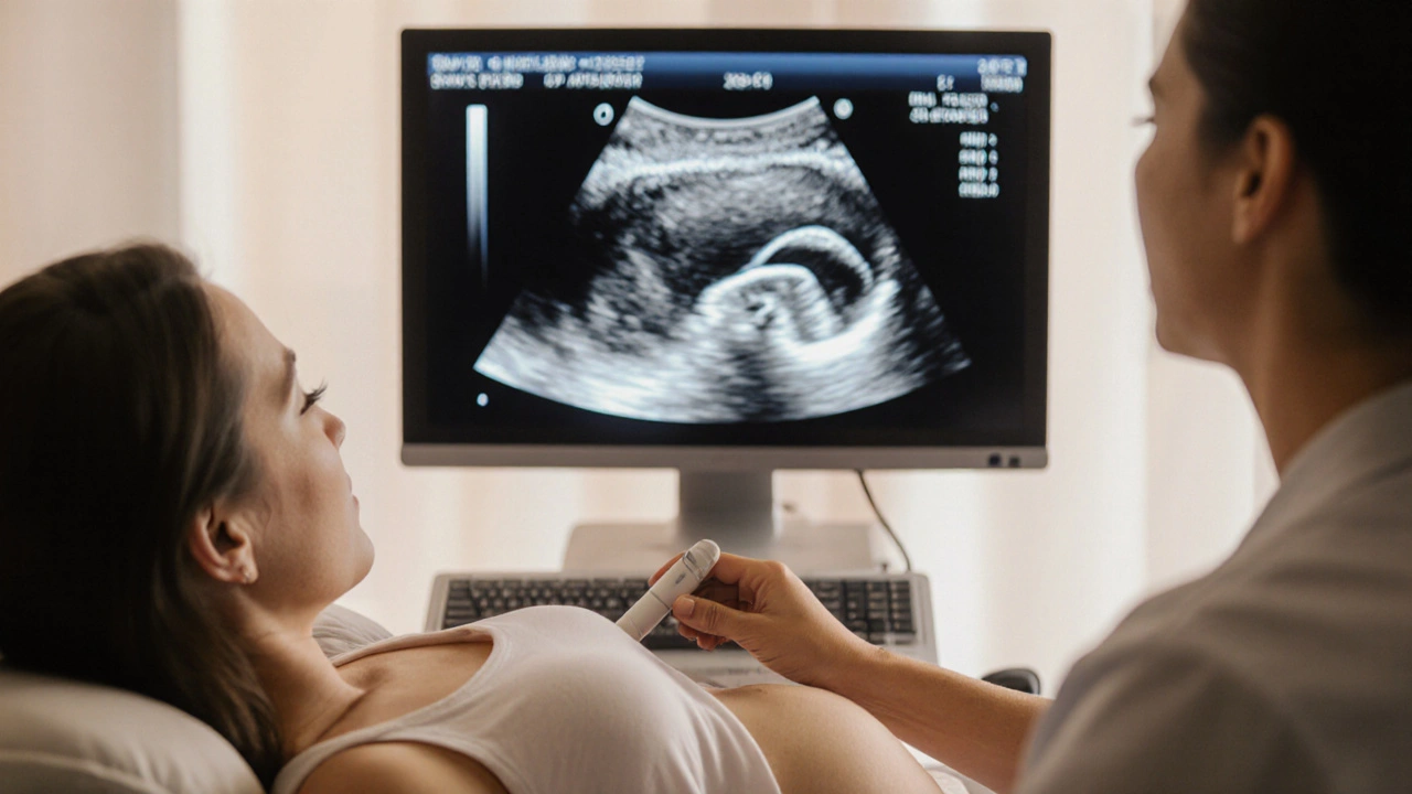
When an expectant mother undergoes a routine scan, Spina bifida is a neural tube defect where the backbone and spinal canal do not close completely, potentially leading to nerve damage can often be spotted long before birth. spina bifida detection relies heavily on the quality of the imaging and the expertise of the sonographer. This article breaks down what spina bifida is, why catching it early matters, and exactly how ultrasound does the heavy lifting.
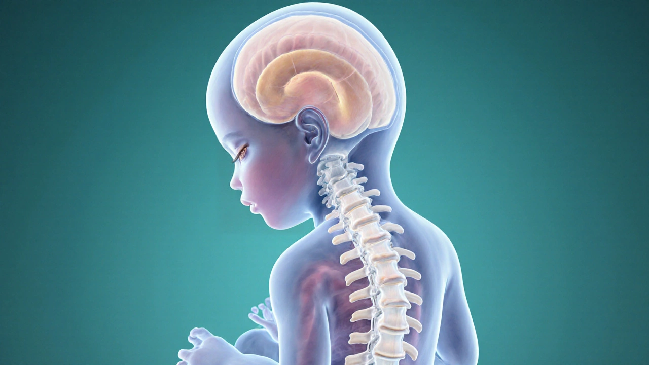
What Is Spina Bifida?
Spina bifida belongs to a broader family called neural tube defects (abnormalities that arise when the embryonic neural tube fails to close properly). The condition ranges from mild (occipital meningomyelocele) to severe (myelomeningocele), with outcomes tied to the level of the spinal opening and associated brain anomalies such as Chiari II malformation.
According to the UK National Health Service, about 1 in 1,000 births involves spina bifida, making it one of the most common congenital disabilities. Early knowledge empowers families to plan surgeries, therapies, and support services well before delivery.
Why Early Detection Changes the Game
Finding spina bifida in the first or early second trimester offers several tangible benefits:
- Medical planning: Fetuses with myelomeningocele may qualify for in‑utero repair, a procedure shown in the MOMS trial to reduce shunt‑dependent hydrocephalus by 50%.
- Psychological preparation: Parents gain time to connect with specialist teams, join support groups, and consider birth‑center options.
- Risk assessment: Some forms of spina bifida are linked with genetic syndromes; early detection prompts targeted genetic testing.
Delaying diagnosis until the third trimester limits surgical options and often forces rushed decision‑making.
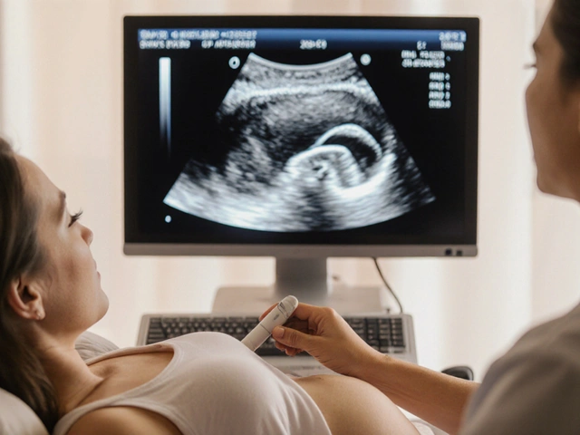
How Ultrasound Spots the Defect
Ultrasound is a non‑invasive imaging method that uses high‑frequency sound waves to create real‑time pictures of fetal anatomy. Modern scanners operate at frequencies between 3 and 8MHz, balancing penetration depth with resolution.
The key sonographic markers for spina bifida include:
- “Lemon sign” - a scalloped frontal bone contour visible as early as 12weeks.
- “Banana sign” - a curved cerebellar shape reflecting posterior fossa crowding.
- Direct visualization of the vertebral arches and spinal cord continuity.
- Associated findings such as ventriculomegaly or spinal fluid leakage.
These signs become clearer as the fetus grows, which is why most protocols schedule a detailed anatomy scan at 18‑20weeks.
Optimal Timing: When to Scan
Two windows dominate clinical practice:
- First‑trimester screening (11‑14weeks): High‑frequency transducers can capture the lemon sign and early spinal defects, though sensitivity hovers around 60%.
- Second‑trimester anatomy scan (18‑22weeks): Sensitivity rises to 95% for myelomeningocele, and the banana sign becomes unmistakable.
If a first‑trimester scan raises concern, a targeted follow‑up at 20weeks is standard. Some centers also perform a third‑trimester check if surgical planning is under consideration.
2D vs. 3D/4D Ultrasound: What’s the Difference?
Traditional 2D ultrasound provides planar images, sufficient for most screening purposes. However, 3D and 4D (real‑time 3D) technologies add volume rendering, allowing clinicians to rotate the fetus and view the spine from multiple angles.
| Feature | 2D Ultrasound | 3D/4D Ultrasound |
|---|---|---|
| Resolution of spinal arches | Good for larger defects | Excellent-allows surface reconstruction |
| Detection of subtle cranial signs | Limited | Improved visualization of lemon/banana signs |
| Utility for surgical planning | Rarely used | High-helps map defect size for fetoscopic repair |
| Cost & availability | Widely available, low cost | Higher cost, limited to tertiary centers |
Most NHS maternity units rely on 2D for routine checks, reserving 3D/4D for cases where a detailed anatomical map is needed.
Complementary Tests: Going Beyond the Scan
Ultrasound rarely works in isolation. The following adjuncts sharpen diagnostic certainty:
- Maternal serum α‑fetoprotein (AFP) (a protein elevated in the maternal blood when the fetus has an open neural tube defect). Elevated AFP at 16‑18weeks can trigger a targeted ultrasound.
- Fetal MRI (magnetic resonance imaging that provides high‑contrast images of fetal brain and spinal structures). MRI is especially useful for assessing associated Chiari II malformation.
- Amniocentesis (sampling of amniotic fluid for genetic analysis and AFP measurement). Offers definitive confirmation but carries a small miscarriage risk (~0.1‑0.3%).
- Chorionic villus sampling (CVS) (early genetic testing at 10‑13weeks via placental tissue). Useful when a hereditary syndrome is suspected.
Integrating these tests with ultrasound findings yields a diagnostic accuracy of >98% for myelomeningocele.
Accuracy, Limitations, and Common Pitfalls
Even the best scanners can miss small lesions. Key challenges include:
- Maternal habitus: High BMI reduces acoustic windows, lowering image clarity.
- Fetal position: Breech or transverse lie may hide the spine during the limited 20‑minute scan window.
- Operator experience: Sonographers with ≥5years of obstetric imaging show a 15% higher detection rate.
- False positives: Isolated lemon signs can appear in healthy fetuses; follow‑up scans are essential.
When uncertainty persists, clinicians typically recommend a repeat scan at 24weeks, possibly supplemented by fetal MRI.
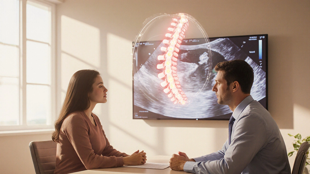
After a Positive Screen: Next Steps for Parents
Receiving a possible spina bifida diagnosis can feel overwhelming. A clear roadmap helps reduce anxiety:
- Consult a maternal‑fetal medicine specialist. They can explain the type of defect, likely outcomes, and surgical options.
- Consider genetic counseling. Some cases link to folate metabolism genes (e.g., MTHFR), influencing future family planning.
- Discuss prenatal surgery. In‑utero repair is offered at select UK centers for myelomeningocele between 19‑26weeks.
- Plan post‑natal care. Early referral to pediatric neurosurgery, physiotherapy, and developmental services improves long‑term quality of life.
- Join support networks. Organizations like Spina Bifida Association UK provide peer mentorship.
Importantly, most families report that having concrete information, even when the news is tough, leads to better emotional coping.
Practical Checklist for Expectant Parents
- Ask when your anatomy scan is scheduled and whether it includes a detailed spine assessment.
- Confirm the clinic uses at least a 2‑MHz transducer for optimal resolution.
- If you have a high BMI, request a longer scan time or a referral to a specialist unit.
- Inquire about the availability of 3D/4D imaging if a defect is suspected.
- Discuss the role of maternal serum AFP and whether you should have the test.
- Know the contact details of the nearest fetal surgery center, should you need it.
Frequently Asked Questions
Can spina bifida be cured after birth?
There is no cure, but surgical repair, physical therapy, and early intervention can greatly improve mobility and independence. The severity of the defect determines the extent of needed support.
How reliable is the lemon sign for early detection?
The lemon sign has a sensitivity of about 70% in the first trimester and a specificity near 95%. It’s a useful flag but always followed by a targeted second‑trimester scan.
Is prenatal surgery safe for the mother?
Maternal complications occur in roughly 10% of cases, most commonly uterine incision issues. The procedure is performed only in specialized centers after thorough risk‑benefit counseling.
What are the chances of having another child with spina bifida?
If a single case occurs without a known genetic cause, recurrence risk is about 2‑4%. With a family history or identified folate‑pathway mutation, the risk can rise to 10% or higher.
Do I need to take extra folic acid if a defect is detected?
High‑dose folic acid (4mg daily) is recommended for the remainder of the pregnancy and for at least three months before trying again, as it reduces the risk of other neural tube defects.


Comments (20)
liza kemala dewi
October 14, 2025 AT 18:02 PMFrom an ethical standpoint, the early identification of neural tube defects such as spina bifida raises profound questions about the responsibilities of both healthcare providers and expectant parents. The principle of beneficence compels clinicians to furnish the most accurate diagnostic information possible, allowing families to make informed decisions regarding prenatal interventions. Autonomy, meanwhile, is respected when parents are presented with comprehensive data about the spectrum of surgical options, potential outcomes, and long‑term care considerations. Moreover, the principle of non‑maleficence underscores the need for precise imaging techniques, because false‑positive findings may lead to unnecessary anxiety or invasive procedures. The integration of maternal serum α‑fetoprotein measurements with high‑resolution ultrasound exemplifies a multimodal approach that mitigates the risk of misdiagnosis. It is also noteworthy that the timing of the ultrasound-first versus second trimester-has implications not only for detection rates but also for the window of opportunity for in‑utero repair, an area that continues to evolve ethically and technically. Beyond the immediate clinical context, public health policies that promote preconception folic acid supplementation have demonstrably reduced the incidence of spina bifida, reflecting a preventive ethic that complements diagnostic excellence. In counseling sessions, it is advisable to discuss the psychosocial support structures available, ranging from specialized fetal therapy centers to peer‑led advocacy groups. Such discourse can empower families, fostering resilience in the face of what may be perceived as daunting prognoses. The nuanced understanding of sonographic markers, such as the lemon and banana signs, should be communicated with appropriate caveats, emphasizing that these signs are part of a broader diagnostic mosaic. Ultimately, the convergence of advanced imaging, biochemical screening, and interdisciplinary consultation creates a robust framework for navigating the complexities inherent in spina bifida detection. By upholding these ethical pillars, clinicians can ensure that the benefits of early detection are realized without compromising the dignity and agency of the families involved. Furthermore, ongoing research into fetal MRI techniques offers the promise of even greater specificity, thereby reinforcing the collaborative ethos between radiology and maternal‑fetal medicine. Continued investment in training sonographers to recognize subtle cranial markers ensures that detection rates improve across diverse healthcare settings. Finally, transparent communication about the limitations of each modality preserves trust and mitigates the potential for misinformation.
Jay Jonas
October 19, 2025 AT 09:09 AMYo, the 3D scan is like magic-suddenly you can see the tiny spine curling around like a pretzel. If you’re lucky enough to get that level of detail, it’s a total game‑changer for the doc’s game plan.
Liam Warren
October 23, 2025 AT 10:22 AMFrom a technocratic perspective, the sensitivity differential between 2D (≈95%) and 3D/4D ultrasound platforms can be attributed to volumetric rendering algorithms that enhance axial resolution. Incorporating Doppler flow metrics further refines the detection of associated ventriculomegaly, which is a critical comorbidity in myelomeningocele cases.
Brian Koehler
October 26, 2025 AT 20:42 PMIndeed-one must commend the obstetric imaging community for its relentless pursuit of diagnostic fidelity; the advent of high‑frequency transducers has undeniably elevated our capacity to visualize minute neuroanatomical anomalies! Moreover, interdisciplinary collaboration between sonographers, radiologists, and fetal surgeons paves the way for integrated care pathways that are nothing short of revolutionary.
Dominique Lemieux
October 29, 2025 AT 18:09 PMWhile the prevailing consensus celebrates early ultrasound screening as a triumph of modern obstetrics, it is imperative to interrogate the underlying assumptions that drive this enthusiasm. The presumption that earlier detection uniformly translates to better outcomes neglects the nuanced realities of healthcare accessibility, socioeconomic disparities, and the psychological burden imposed upon expectant families. For instance, in regions where specialized fetal surgery centers are scarce, a first‑trimester diagnosis may simply generate anxiety without offering actionable therapeutic avenues. Additionally, the reliance on sonographic markers such as the lemon sign, albeit statistically robust, is not infallible; false positives linger in the literature, occasionally precipitating invasive follow‑up procedures that carry their own risk profiles. Therefore, a balanced appraisal demands that we integrate epidemiological data with a sober appraisal of systemic limitations before heralding early detection as an unqualified boon.
Laura MacEachern
November 1, 2025 AT 01:42 AMHigh BMI can really blur those crucial images.
BJ Anderson
November 3, 2025 AT 03:42 AMThe moment the ultrasound shows that unmistakable banana sign, it feels like a spotlight has been switched on, illuminating a path that previously lay shrouded in uncertainty. Families suddenly confront a cascade of decisions-whether to pursue in‑utero repair, how to prepare for neonatal care, and what long‑term support will look like. The gravity of that instant is both terrifying and empowering, a duality that underscores the profound impact of prenatal imaging.
Alexander Rodriguez
November 5, 2025 AT 00:09 AMEarly scans catch most cases, especially after week 18, and that helps doctors plan surgery and give parents clear info.
Abhinav Sharma
November 6, 2025 AT 15:02 PMIt’s fascinating how the integration of serum AFP levels with targeted ultrasound can refine diagnostic accuracy-essentially turning a probability problem into a near‑certainty. This synergy exemplifies precision medicine in prenatal care 😊.
Welcher Saltsman
November 8, 2025 AT 00:22 AMjust a heads up if you’re over 30 BMI ask for a longer scan it really helps get clear pics
april wang
November 9, 2025 AT 04:09 AMBuilding upon the technical nuances you highlighted, it is worth exploring how the evolution of probe technology-specifically the shift from curvilinear to micro‑convex arrays-has substantially mitigated acoustic shadowing, thereby enhancing the visualization of subtle vertebral arch defects. The implementation of speckle reduction filters further refines image clarity, allowing clinicians to discern the faint contours of the spinal cord amidst surrounding fluid. Moreover, advances in machine learning algorithms are now being leveraged to automatically flag anomalous patterns, reducing observer variability and expediting diagnostic workflows. In practice, these innovations translate to a higher confidence level when interpreting the lemon and banana signs, especially in cases where fetal positioning is suboptimal. Coupled with standardized training modules, the proficiency of sonographers across diverse practice settings can be elevated, diminishing the disparity in detection rates between high‑resource and low‑resource environments. Ultimately, the confluence of hardware enhancements, software sophistication, and targeted education forms a synergistic triad that propels the field toward more reliable and equitable prenatal diagnostics.
Franco WR
November 10, 2025 AT 05:09 AMEchoing the ethical framework you articulated, I would like to underscore the pivotal role of informed consent in the context of prenatal imaging. When couples are presented with probabilistic data-such as a 70% sensitivity for first‑trimester lemon sign detection-they must also be apprised of the residual uncertainty inherent in any screening modality. Transparent communication about the potential need for confirmatory testing, be it targeted ultrasound or fetal MRI, safeguards against the inadvertent erosion of trust that can arise from ambiguous results. Furthermore, cultural considerations often influence how families perceive risk and intervention, necessitating a culturally competent approach that respects diverse belief systems while delivering evidence‑based guidance. The integration of multidisciplinary teams-including genetic counselors, neonatologists, and psychosocial support specialists-ensures that families receive holistic care that transcends purely medical advice. By embedding these principles into routine practice, we reinforce the notion that early detection is not merely a technical achievement but a compassionate endeavor rooted in respect for patient autonomy and dignity.
Rachelle Dodge
November 11, 2025 AT 03:22 AMThe lemon sign’s early reveal is like nature’s neon warning light, flashing bright enough to catch any watchful eye.
Gaurav Joshi
November 11, 2025 AT 22:49 PMIn addition to the imaging modalities discussed, it’s essential to recognize that maternal nutrition, particularly folic acid intake, remains a cornerstone in preventing neural tube defects. While the technology offers remarkable detection capabilities, primary prevention can reduce the overall incidence, thereby lessening the reliance on complex prenatal interventions.
Elaine Proffitt
November 12, 2025 AT 15:29 PMAccurate timing of the anatomy scan matters.
Christopher Munt
November 13, 2025 AT 05:22 AM2D scans are good enough for most cases 👍
Sangeeta Birdi
November 13, 2025 AT 16:29 PMHaving gone through the process myself, I can say that a supportive fetal medicine team makes all the difference 😊. When the ultrasound tech takes extra time to adjust the probe and explain each view, it eases the anxiety that often accompanies a tentative diagnosis. The combination of clear imaging and compassionate counseling turns a potentially overwhelming experience into an empowering one.
Chelsea Caterer
November 14, 2025 AT 00:49 AMThs infro rmation is vry helpful for future parents.
Lauren Carlton
November 14, 2025 AT 06:22 AMThe article contains numerous comma splices and inconsistent tense usage, which detracts from its credibility.
Katelyn Johnson
November 14, 2025 AT 09:09 AMOverall the post gives a solid overview of ultrasound use for spina bifida detection